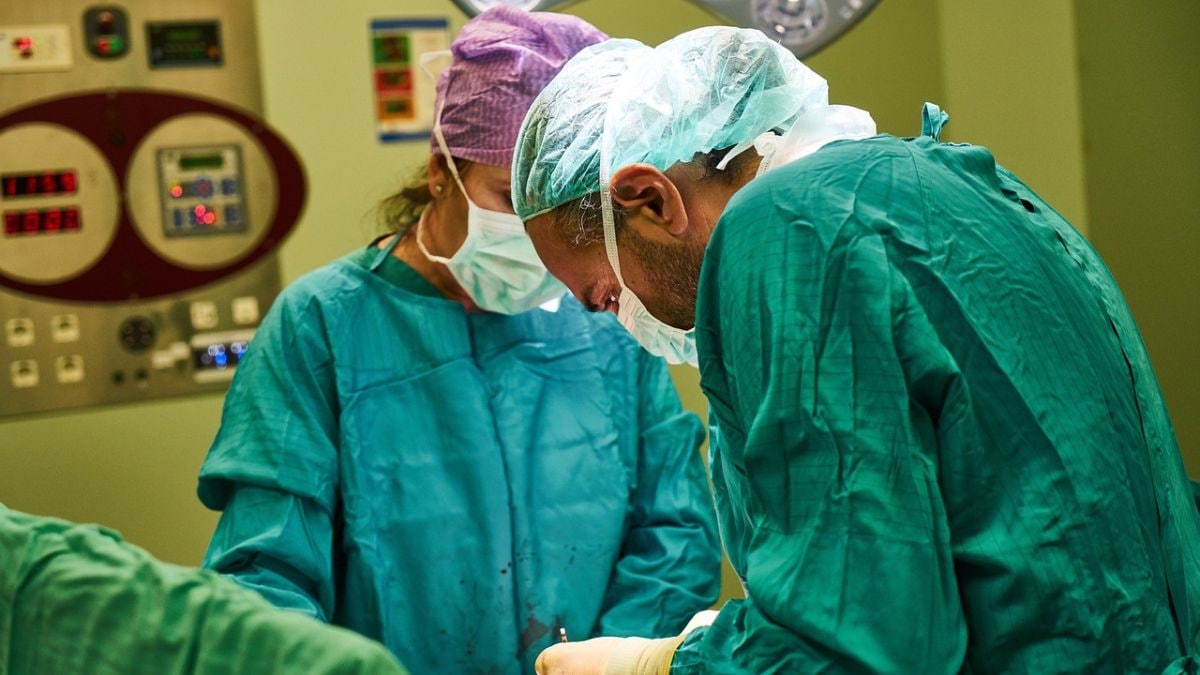
A groundbreaking artificial intelligence tool called FastGlioma has been developed, enabling surgeons to detect residual cancerous brain tumors within 10 seconds during surgery. The innovation, detailed in a recent study in Nature, is seen as a significant advancement in neurosurgery, outperforming traditional tumor detection methods. Researchers from the University of Michigan and the University of California, San Francisco, led the study, highlighting its potential to improve surgical outcomes for patients with diffuse gliomas.
Todd Hollon, MD, a neurosurgeon at the University of Michigan Health, described FastGlioma as a transformative diagnostic tool that provides a faster and more accurate method for identifying tumor remnants. He noted its ability to reduce reliance on current methods, such as intraoperative MRI or fluorescent imaging agents, which are often inaccessible or unsuitable for all tumor types.
Addressing Residual Tumors During Surgery
As per the study from Michigan Medicine – University of Michigan, residual tumors, which often resemble healthy brain tissue, are a common challenge in neurosurgery. Surgeons have traditionally struggled to differentiate between healthy brain and remaining cancerous tissue, leading to incomplete tumor removal. FastGlioma addresses this by combining high-resolution optical imaging with artificial intelligence to identify tumor infiltration rapidly and accurately.
In an international study, the model was tested on specimens from 220 patients with low- or high-grade diffuse gliomas. FastGlioma achieved an average accuracy of 92%, significantly outperforming conventional methods, which had a higher miss rate for high-risk tumor remnants. Co-senior author Shawn Hervey-Jumper, MD, professor of neurosurgery at UCSF, emphasized its ability to enhance surgical precision while minimizing the dependence on imaging agents or time-consuming procedures.
Future Applications in Cancer Surgery
FastGlioma is based on foundation models, a type of AI trained on vast datasets, allowing adaptation across various tasks. The model has shown potential for application in other cancers, including lung, prostate, and breast tumors, without requiring extensive retraining.
Aditya S. Pandey, MD, chair of neurosurgery at the University of Michigan, affirmed its role in improving surgical outcomes globally, aligning with recommendations to integrate AI into cancer surgery. Researchers aim to expand its use to additional tumor types, potentially reshaping cancer treatment approaches worldwide.


